Pupillary Evaluation for Differential Diagnosis of Coma

Pupil examination is a simple and non-invasive test that may provide valuable information about the health of our vision and nervous system. It is necessary to measure pupil size, equality, and responsiveness because changes in the pupils’ size, equality, and reactivity might give valuable diagnostic information. Using this article, we want to shed some insight into the fundamental approaches of pupillary examination and how they might be utilized to diagnose coma and neurological disorders.
What is the pupil?
The pupil is a small aperture in the center of the iris that enables light to enter the retina. It is placed in the center of the iris. The quantity of light that enters the eye is determined by the size of the pupil of the eye. The iris’s dilator and sphincter muscles are responsible for controlling the size of the pupil. There is usually just one practically central pupil (slightly nasal), and the size of the pupil fluctuates from 2.5 to 4 mm depending on the amount of light present.
What is a coma?
A coma is nothing but a condition of unconsciousness that lasts for an extended period of time. During a coma, a person is entirely unaware of his or her surroundings. The individual seems to be awake yet appears to be asleep. However, unlike in a deep slumber, the individual cannot be roused from their sleep by any external stimuli, such as pain.
What Causes a Coma?
Comas are induced by a traumatic brain injury (TBI). Increased pressure, hemorrhage, oxygen deprivation, or toxic accumulation in the brain is all possible causes of brain damage. The harm may be transitory and reversible. It is also possible that it will be permanent. According to research, over 50% of comas are caused by head trauma or disruptions in the brain’s circulatory system.
Glasgow Coma Scale
A head injury may result in severe brain damage, and the Glasgow Coma Scale (GCS) can be used to specify the stretch of the damage.
Patient reactions, including verbal and physical replies and how easy they can open their eyes, are weighed to get a score.
Eyes: Scores range from 1 to 4, with one indicating that a person does not open their eyes, two indicating that they open their eyes in reaction to pain, three indicating that they open their eyes in response to the speech, and four indicating that they open their eyes on their initiative.
A score of 1 indicates that the individual does not make any sound, two signifies that they mutter but cannot be heard, three indicates that they utter unsuitable phrases, four indicates that they talk but are confused, and five indicates that the person communicates normally.
Response to pain is measured on a scale of 1 to 6, with 1 to 5 indicating the person’s response to physical discomfort. One score represents no movement, two signifies that they straighten a limb in reaction to pain, three signifies that they respond unusually to pain, four represents that they move away from discomfort, and five represents that they can locate the source of their suffering. A score of 6 indicates that the individual may be trusted to follow instructions.
A total score of 8 or fewer implies that the person is in a coma. If the score falls between 9 and 12, the condition is considered moderate. Generally speaking, if the score is 13 or more, the damage to awareness is mild.
What actually occurs in the brain during a coma?
Take on the perspective of a comatose individual. A coma may be caused by a variety of things, including a severe brain injury, a stroke, or even a near-drowning incident. The comatose patient lies quietly on the bed, eyes closed. The individual does not display any evidence of communication with the surroundings. However, the comatose individual does not react and seems to be apathetic to what is going on around them. A person is said to be unconscious while they are in a coma. Is a coma patient’s brain still functioning?
If a person is senseless, it is possible that their brain is still able to interpret information from the outer world, such as the sound of your voice when you talk to them or the footfall of someone coming. A technique known as electroencephalography (EEG) is used to monitor the brain activity of a person who is in a coma. Electrodes are placed on the patient’s head to record the electrical activity of the brain. Electroencephalography (EEG) is a tool that may help us monitor the activity of brain cells known as neurons. Placing electrodes on a person’s head allows us to track their brain’s electrical activity. Caps hold these electrodes. To visualize this, consider a swimming hat with several punctures.
How can pupil measurement and evaluation help in the diagnosis and management of coma?
Pre- and post-exposure pupil diameter measurement of the three main characteristics of the eye’s pupils is part of the process of doing a comprehensive examination of the eyes. In most people, the diameter of the pupils is between 2 and 6 millimeters, although they may be as big as 9 millimeters in certain people. Pupils may also be pinpoint, tiny, big, or dilated in any of these ways. The form of a normal pupil is round, but it may also be uneven, keyhole-shaped, or even oval.
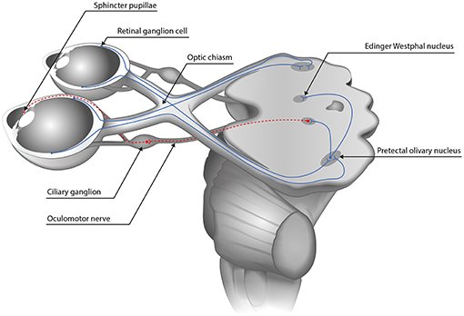
With both eyes open, the doctor will shine a light into each patient’s eyes in order to determine the size of the pupil. Right away, both eyes should contract. When they remove the light from one eye, the other will dilate instantaneously. We’ll note the speed, sluggishness, lack of reaction, and stability of the answer. Report any deviations from the norm as soon as you see them. Pupils may dilate or dilate unevenly as a consequence of developing a neurologic disease in many circumstances.
Pupils that are fixed and dilated should be reported to a physician right away (unless the patient’s pupils have recently been chemically dilated). You might anticipate your doctor to request further tests like a CT scan if you see significant changes in your pupillary response.
Conclusion:
NeurOptics is well-equipped to handle all pupil measurements and issues related to the eye. And with our cutting-edge pupillometer, the NPi®-300, any eye problem you may have will be detected quickly and the appropriate treatment given.


